Sublethal concentrations of dichlorvos and paraquat induce genotoxic and histological effects in the Clarias gariepinus
Article information

Abstract
Non-target aquatic organisms such as fish may be impacted by agricultural activities through the run-off of pesticides from farms into aquatic ecosystems. In this study, the genotoxic (erythrocytic micronuclei) and histological effects of sublethal concentrations (1% and 10% of 96-h median lethal concentration (LC50) values) of two pesticides (dichlorvos and paraquat) were evaluated in Clarias gariepinus (the African Sharptooth Catfish) for 28 days. The 96-h LC50 of dichlorvos and paraquat against fingerlings of C. gariepinus was 730 μg/L and 50 μg/L, respectively. There was a significant dose-dependent increase (p<0.05) in micronuclei in the erythrocytes of exposed C. gariepinus (2.00±0.82 ‰ to 3.25±1.26 ‰ for dichlorvos and 2.25±0.96 ‰ to 4.75±0.96 ‰ for paraquat) compared to control (0.75±0.96 ‰) by day 28. Gill histological alterations such as mild to severe necrosis and blunting of secondary lamellae were observed in C. gariepinus exposed to higher sublethal concentrations of both pesticides. This study showed that non-target aquatic organisms like C. gariepinus may be at risk of adverse biological effects from exposure to pesticides from non-point sources. We recommend environmental monitoring and sensitization on responsible pesticide use to stakeholders. This will forestall potential adverse ecological effects in aquatic ecosystems.
Introduction
The increase in global population has resulted in a teeming demand for increased food crop production which is aided through the use of pesticides [1]. Consequently, various classes of pesticides targeting specific pests are being produced to promote increased and quality crop yields. Dichlorvos (2,2-dichlorovinyl dimethyl phosphate) is one of the most commonly used organophosphate pesticide with insecticidal properties and often applied on crops, stored products and in treatment of animals [2,3]. It has been used to eliminate crustacean ectoparasites in fish farming [4]. Paraquat (1,1′-dimethyl-4-4′-bipyridinium) and its dichloride salt (1,1′-dimethyl-4-4′-bipyridinium dichloride) are one of the most widely used herbicides for controlling weeds especially in developing countries [5]. Further, it is applied for defoliating and desiccating purposes in the harvest of economic crops such as cotton, beans, soybeans, potatoes, sunflowers, sugarcanes, among others [6]. The ubiquity of these pesticides in various environmental matrices including surface waters is due to their relatively low cost, high availability and high solubility in water [7–9]. These pesticides reach the aquatic ecosystem through various routes such as direct application, spray-drift, run-off, leaching, and discharge from factories and sewage [10].
Pesticides use has increased the exposure of organisms inhabiting the aquatic environment [11] causing hazardous effects to non-target organisms [12], disrupting the food chain, modifying the food web and causing instability in the ecosystem [13]. Biomarkers such as micronuclei (MN) induction indicating genotoxic effects and histopathological changes have been utilized as indices of toxicant exposure or effect in Clarias gariepinus [14,15], other fish species [16,17] and animals [18]. Fragmentation and/or MN induction results from alterations in erythrocyte nuclei [19] which could lead to growth retardation, abnormal development and deleterious effects at population and community levels [17,20]. Further, studies to elucidate the toxic effects of dichlorvos and paraquat in C. gariepinus include acute toxicity of dichlorvos [21,22] and paraquat [23,24], hepatocyte vacuolation in C. gariepinus exposed to acute levels of paraquat [25]. However, few studies have evaluated the genotoxic and histological effects of dichlorvos and paraquat at sublethal or environmentally relevant levels in C. gariepinus.
The African sharptooth catfish, C. gariepinus is a highly economical and ecologically relevant fish species [26]. It belongs to the family Clariidae and is a common model fish species utilized for aquatic toxicological studies among other animal experimental studies in Nigeria [27–30]. The aim of this study was to evaluate the effects of sublethal (environmentally relevant) concentrations of two pesticides (dichlorvos and paraquat) in C. gariepinus for 28 days. The results will provide information on the potential effects of these frequently used pesticides particularly in areas that are close to surface waters.
Materials and Methods
Test Pesticides
The pesticides used in this study were dichlorvos (DAKSH – active ingredient 100% Emulsifiable Concentrate in 1 L), and paraquat (Paraforce – active ingredient 200 g W.C. in 1 L). They were obtained from a commercial vendor in Lagos, Nigeria. The pesticides were in liquid form and soluble in water. Stock solutions were prepared of 1 mL/L of each pesticide. Hence, stock solution of paraquat was 200 mg/L.
Test animal procurement and acclimatization
Fingerlings (weight range: 7–11 g; length range: 5.0–6.5 cm) and juveniles (weight range: 16.3–30 g; length range: 14.5–18.3 cm) of C. gariepinus (Chordata, Osteichthyes, Siluriformes, Clariidae) were obtained from a local aquaculture farm in Bariga, Lagos, Nigeria. The fish were kept in different 60 L holding tanks half-filled with dechlorinated water and allowed to acclimatize to laboratory conditions (temperature: 28±2 °C; relative humidity: 78±4%) for a week prior to tests. The fish were fed twice daily with commercial Coppens fish feed (size: 1–3 mm) and water was changed daily in the holding tanks to prevent accumulation of toxic waste substances. Feeding was stopped 24 h prior to exposure of fish to test chemicals [31].
Acute toxicity studies with Clarias gariepinus exposed to dichlorvos and paraquat
The acute toxicity studies were conducted using C. gariepinus following the OECD (the organization for economic co-operation and development) protocol [32] for fish acute toxicity testing. The bioassay was conducted using plastic tanks measuring 5 L covered with a mesh to prevent the escape of the fish. Five fingerlings were selected randomly into the test tanks for toxicity testing. After a range finding test, the fingerlings were exposed to varying concentrations; 200 μg/L, 400 μg/L, 700 μg/L, 1000 μg/L, 1500 μg/L and control (dechlorinated water alone) for dichlorvos and 15 μg/L, 30 μg/L, 44 μg/L, 88 μg/L, 175 μg/L and control (dechlorinated water alone) for paraquat. Fish were considered dead when there was no movement and the body was floating horizontally at the top or sinking at the bottom of the test media. Dead fish were removed and disposed immediately to prevent contamination of the test media [31].
Experimental design for sublethal toxicity studies with Clarias gariepinus exposed to dichlorvos and paraquat
The sublethal toxicity studies with dichlorvos and paraquat on juveniles of C. gariepinus were carried out for a period of 28 days. The fish were kept in plastic containers measuring 5 L covered with mesh to prevent the escape of the fish. Five juveniles were randomly selected for the sublethal studies. The 10% and 1% of the 96-h median lethal concentration (LC50) values of dichlorvos and paraquat against the fingerlings of C. gariepinus were used. The juveniles were exposed to 73 μg/L (10% 96-h LC50), 7.3 μg/L (1% 96-h LC50) for dichlorvos and 5 μg/L (10% 96-h LC50), 0.5 μg/L (1% 96-h LC50) for paraquat and control (water alone). A semi-static bioassay protocol was followed in which the test media was changed every 48 hours to prevent contamination of the test media with waste metabolites [31,32].
Genotoxicity studies (Micronucleus assay) of dichlorvos and paraquat against Clarias gariepinus
At 28-day post-exposure, two active juveniles were randomly selected from each treatment, blood was drawn from the caudal vein with a 2 mL syringe, smeared on a clean glass slide at an angle of 45° and air dried overnight at room temperature. The slides were fixed in absolute methanol for five mins and air dried. Subsequent staining was conducted using May-Grunwald Giemsa stain. The smears were observed under a light microscope (Leica® DM500, Wetzlar, Germany) with an objective lens of ×100 and scored for MN [15].
Histological studies of dichlorvos and paraquat against Clarias gariepinus
At 28-day post-exposure, two active juveniles from which blood was drawn (for micronucleus assay) were euthanized and dissected. The gill and liver from each fish were harvested and prepared for histological sections. The organs were fixed in Bouin’s fluid and subsequently dehydrated through a graded series of ethanol [18]. Following this, they were embedded in paraffin and then manually sectioned with a microtome at 4–5 μm. The sections were dewaxed and stained with hematoxylin and eosin (H&E) and examined using a digital light microscope (Leica® DM500) [31].
Statistical analysis
The toxicological dose-response data involving quantal response (mortality) for acute toxicity studies was calculated using the probit analysis (Finney 1971) to derive the median lethal concentrations (concentration of test pesticides that causes 50% mortality of exposed test fishes) over a period of 96-h (96-h LC50). Toxicity factor (TF) was calculated as the ratio of the least toxic pesticide to the most toxic pesticide. The analysis was conducted using IBM SPSS version 20.0. The micronucleus assay results are presented as mean±standard error. One-way analysis of variance (ANOVA) was used to test for differences between sublethal concentrations of the test pesticides and control. Significant differences between the treatment means were analyzed with Tukey HSD posthoc test with the level of significance set at p<0.05. The MN assay data analysis was conducted using IBM SPSS version 20.0.
Results
Acute toxicity studies of dichlorvos and paraquat against Clarias gariepinus fingerlings
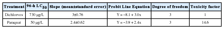
The 96-h LC50 values of dichlorvos and paraquat against fingerlings of C. gariepinus were 730 μg/L and 50 μg/L respectively (Table 1). The computed toxicity factor showed that paraquat was approximately 15 times more toxic than dichlorvos (Table 1).
Relative acute toxicity of dichlorvos and paraquat against Clarias gariepinus fingerlings
Genotoxicity (micronuclei induction) of sublethal concentrations of dichlorvos and paraquat in erythrocytes of Clarias gariepinus juveniles
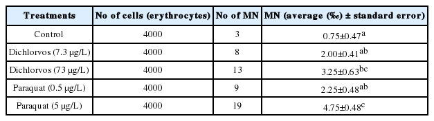
A significantly higher (p<0.05) induction of MN was observed in the erythrocytes of C. gariepinus exposed to the higher sublethal concentrations of the pesticides, dichlorvos 73 μg/L (3.25±0.63‰) and paraquat 5 μg/L (4.75±0.48‰) compared to the control (0.75 ± 0.47 ‰) (Table 2). There was a non-significant (p>0.05) increase in MN induction in C. gariepinus exposed to the higher concentration (73 μg/L) compared to the lower concentration (7.3 μg/L) of dichlorvos (Table 2). On the other hand, a significant (p<0.05) increase in MN induction in C. gariepinus exposed to the higher concentration (5 μg/L) compared to the lower concentration (0.5 μg/L) of paraquat was observed (Table 2).
Micronuclei (MN) in erythrocytes of Clarias gariepinus juveniles exposed to sublethal concentrations of dichlorvos and paraquat at day 28
Histological effects of sublethal concentrations of dichlorvos and paraquat in Clarias gariepinus juveniles
There were no histological alterations in the control fishes evidenced by the preservation of cartilaginous support of the primary lamellae of the gills (Figure 1a and 1b) and parallel radially arranged plates of hepatocytes (Figures 2a and 2b). In dichlorvos- and paraquat-treated C. gariepinus, mild to severe lamellar necrosis in the gills were observed in the highest sublethal concentrations (73 μg/L and 5 μg/L, respectively) (Figure 1c and 1d; Figure 1g and 1h)) while no abnormalities were observed in the lower sublethal concentration (7.3 μg/L) (Figure 1e and 1f; Figure 1i and 1j). No abnormalities were observed in the liver (Figure 2c, 2d and 2e; Figure 2g–j) except for cytoplasmic fat vacuoles in the lowest sublethal concentration of dichlorvos (Figure 2f).
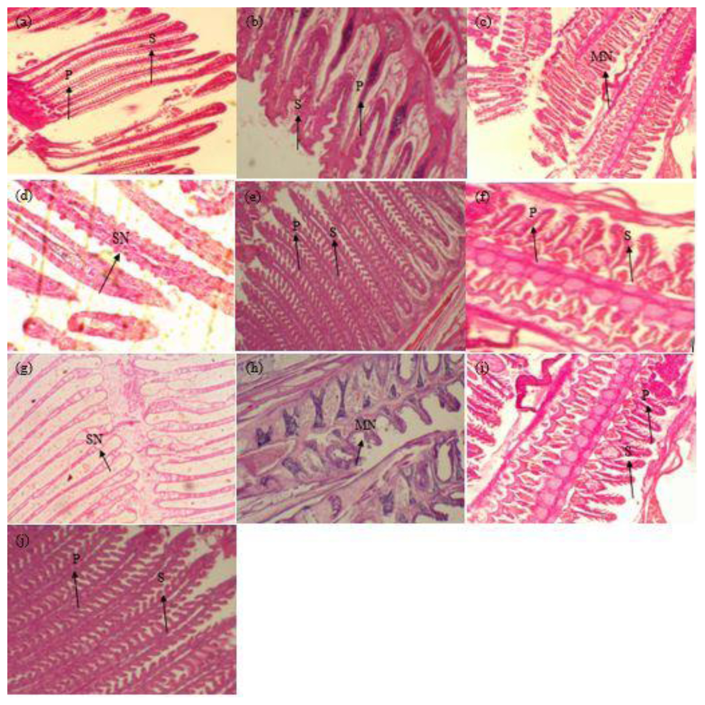
Photomicrograph of histological sections through the gills of Clarias gariepinus exposed to sublethal concentrations of dichlorvos and paraquat over a period of 28 days. (a,b) Controls A and B - Histologic section of gill filament shows preservation of the cartilaginous support of the primary lamellae, with long primary lamellae and comb-like secondary lamellae projecting from both sides of each primary lamella indicating normal gill (X100); (c) Dichlorvos (73 μg/L) A - Histologic sections of gills show shortening/blunting of secondary lamellae but preservation of the primary lamellae indicating mild lamellar necrosis (X100); (d) Dichlorvos (73 μg/L) B - Histologic sections of tissue show necrosis and destruction of both primary and secondary lamellae indicating severe lamellar necrosis (X400); (e) Dichlorvos (7.3 μg/L) A - Histologic section of gill filament shows preservation of the cartilaginous support of the primary lamellae, with long primary lamellae and comb-like secondary lamellae projecting from both sides of each primary lamella indicating normal gill (X100); (f) Dichlorvos (7.3 μg/L) B - Histologic sections of gills show presence of primary and secondary lamellae. No areas of necrosis or inflammation are seen indicating normal gill (X400); (g) Paraquat (5 μg/L) A - Histologic sections of tissue show necrosis and destruction of both primary and secondary lamellae indicating severe lamellar necrosis (X400); (h) Paraquat (5 μg/L) B - Histologic sections of gills show shortening/blunting of secondary lamellae but preservation of the primary lamellae indicating mild lamellar necrosis (X400); (i) Paraquat (0.5 μg/L) A - Histologic section of gill filament shows preservation of the cartilaginous support of the primary lamellae, with long primary lamellae and comb-like secondary lamellae projecting from both sides of each primary lamella indicating normal gill (X400); (j) Paraquat (0.5 μg/L) B - Histologic sections of gills show presence of primary and secondary lamellae. No areas of necrosis or inflammation are seen indicating normal gill (X100).
NOTES: P = Primary gill filaments; S = Secondary lamellae; MN = Mild Necrosis; SN = Severe Necrosis
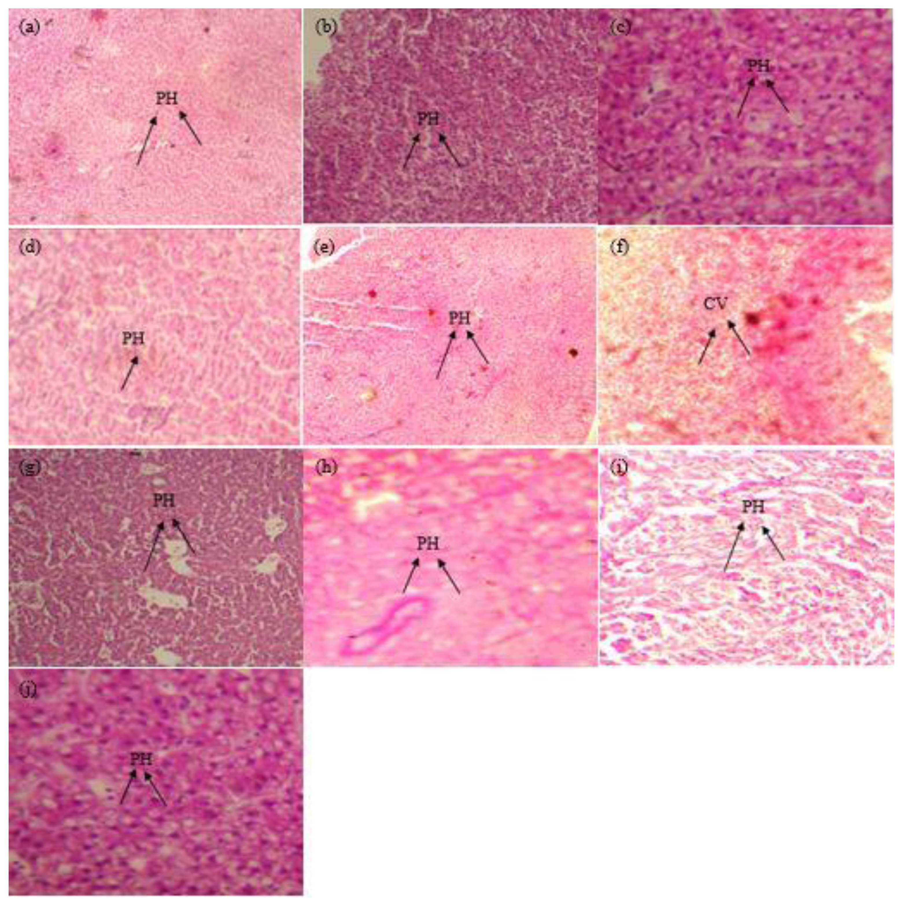
Photomicrograph of histological sections through the liver of Clarias gariepinus exposed to sublethal concentrations of dichlorvos and paraquat over a period of 28 days: (a,b) Control A,B - Histologic sections of liver tissue show parallel radially arranged plates of hepatocytes. No abnormalities are seen indicating normal liver (X100); (c) Dichlorvos (73 μg/L) A - Histologic sections of liver tissue show parallel radially arranged plates of hepatocytes. No abnormalities are seen indicating normal liver (X400); (d) Dichlorvos (73 μg/L) B-Histologic sections of liver tissue show parallel radially arranged plates of hepatocytes. No abnormalities are seen indicating normal liver (X400); (e) Dichlorvos (7.3 μg/L) A - Histologic sections of liver tissue show parallel radially arranged plates of hepatocytes. No abnormalities are seen indicating normal liver (X400); (f) Dichlorvos (7.3 μg/L) B - Histologic sections of tissue show hepatocytes arranged radially as plates. Cytoplasmic fat vacuoles are seen indicating fatty liver (X400); (g) Paraquat (5 μg/L) A - Histologic sections of liver tissue show parallel radially arranged plates of hepatocytes. No abnormalities are seen indicating normal liver (X100); (h) Paraquat (5 μg/L) B - Histologic sections of liver tissue show parallel radially arranged plates of hepatocytes. No abnormalities are seen indicating normal liver (X100); (i) Paraquat (0.5 μg/L) A - Histologic sections of liver tissue show parallel radially arranged plates of hepatocytes. No abnormalities are seen indicating normal liver (X100); (j) Paraquat (0.5 μg/L) B - Histologic sections of liver tissue show parallel radially arranged plates of hepatocytes. No abnormalities are seen indicating normal liver (X100).
NOTES: PH = Plates of Hepatocytes, CV = Cytoplasmic Fat
Discussion
The evaluation of pesticides’ risk to non-target organisms like fish is imperative to ensure the sustainability of aquatic life from inadvertent exposure to contaminants and prevent human health effects from the consumption of contaminated fish. The observed LC50 values of dichlorvos and paraquat against C. gariepinus in this study varies from the results of previous studies which revealed varying 96-h LC50 values of 275.2 μg/L for fingerlings and 492 μg/L for juveniles [21], 1290 μg/L [22] in C. gariepinus exposed to dichlorvos. Similarly, the results in this study for paraquat varies with the findings of Ayanda et al. [24] – 70 μg/L and Nwani et al. [23] – 27.46 mg/L. These differential responses may be attributed to the formulation of the pesticides used in the studies, the age of species used, time and conditions of exposure. There is a dearth of comparative studies of dichlorvos and paraquat toxicity in fish species hence this current study may provide a basis for comparative studies of the pesticides in other fish species. The differential toxicities observed may be due to their different modes of action.
The dose-dependent increase in the number of MN in the erythrocytes of the exposed fishes in this study is consistent with the findings of Mumuni and Sogbanmu [15] who reported similar observations in C. gariepinus exposed to a mixture of endosulfan and deltamethrin. Similarly, increased MN rate was reported in loach (Misgurnus anguillicaudatus) with the increase in dichlorvos concentrations and treatment time [33]. MN (≤⅓ cell nucleus) formation occurs due to the non-incorporation of a chromosome or its fragment into one of the daughter nuclei during cell division which relates to chromosome aberration [33]. Chromosomal aberrations are induced by clastogenic and aneugenic agents and the MN assay is often used to evaluate the structural and numerical chromosome aberrations [34]. Furthermore, our findings corroborate the observations by Yin et al. [35] who observed significant (p<0.05) concentration-dependent increase in DNA damage using the comet assay in erythrocytes of Chinese toad (Bufo bufo gargarizans) tadpoles exposed to sublethal concentrations of paraquat. The observed increase in MN frequency with increasing test concentration of the pesticides in this study suggests progressive damage to the DNA in C. gariepinus expressed in the chromosome aberration.
Fanta et al. [36] observed that alterations in the organs are normally easier to identify than functional ones and serve as warning signs to the health of an animal. Histopathological studies are acknowledged as a sensitive endpoint for detecting organ toxicity during exposure to toxicants [37] and capable of providing specific information on the acute and chronic effects of toxicants on targeted organs that may not be detected by functional biomarkers [38]. The observed histopathological abnormalities particularly in the higher sublethal concentrations of both pesticides agree with the observations in the MN assay above which can serve as early warning indices for the evaluation of fish health in the aquatic environment.
Conclusion
The study shows that non-target organisms in aquatic ecosystems such as C. gariepinus may be at risk of toxic effects of environmentally relevant (sublethal) concentrations of dichlorvos and paraquat from point and/or non-point sources. Consistent biomonitoring and sensitization of stakeholders on responsible pesticide use are therefore imperative to forestall adverse ecological effects in aquatic ecosystems. Further, ecological risk indices for these pesticides should be developed to safeguard aquatic life within the ambits of the United Nations Sustainable Development Goal 14 (sustaining life below water).
Acknowledgement
The authors gratefully acknowledge the Prof. Joseph K. Saliu (Department of Zoology, Faculty of Science, University of Lagos) and Prof. A. A. Bakare (Department of Zoology, Faculty of Science, University of Ibadan) for their critical evaluations and corrections of the thesis from which this manuscript was culled.
Notes
Ethics Statement
All applicable international, national, and/or institutional guidelines for the care and use of animals were followed. This study followed the principles in the Declaration of Helsinki on the humane treatment of animals used in research (http://www.wma.net/en/30publications/10policies/a18/) and the principles in the AVMA Guidelines for the euthanasia of animals [39].
Conflict of interest
The authors declare that they have no conflict of interest.
CRediT author statement
TOS: Conceptualization, Methodology, Supervision, Writing-original draft preparation, Writing - Reviewing and Editing; EIO: Data curation and analysis, investigation, Writing-original draft preparation; JCA: Co-conceptualization, Writing - Reviewing and Editing.